

Chandra Prakash Dixit1 and Abhishek shokeen2
1Assistant Professor, Department of Veterinary Gynaecology and Obstetrics, Apollo College of Veterinary Medicine, Jaipur, Rajasthan
2Undergraduate student, Apollo College of Veterinary Medicine, Jaipur, Rajasthan
ABSTRACT
A three year old male Pariah dog was presented to the ambulatory unit of Apollo College of Veterinary Medicine, Jaipur, Rajasthan with the history of discomfort and swollen penis. The clinical examination revealed entrapment of the prepuce at the base of the penis (bulbus glandis) with severe swelling. There was slight ulceration in penile tissue, congestion of the penile and preputial tissue. After treatment, the edematous swelling subsided and the animal was recovered uneventfully with no recurrence up to three weeks of follow up.
Keywords: Paraphimosis, Caster sugar, Dog
Introduction
Paraphimosis is the inability to reduce the penis in the preputial cavity, which mainly occurs in dogs due to entrapment and inversion of the preputial tissue behind the bulbus glandis (Boden, 2005). It can occur due to trauma, post coitus, manual semen collection, other causes are priapism, phimosis and chronic irritation (Roberts, 1999; Fossum, 2007). In paraphimosis, the prepuce gets inverted, causes entrapment of the extruded penis and as a sequlae, it causes impairment of the venous drainage and congestion of the penile tissue (Davidson, 2020). Necrosis of penile tissue is very common, if prompt veterinary intervention is not provided. The extruded penis gets swollen due to the impaired venous drainage while due to the external exposure the mucosa becomes dried and inflammed. Self mutilation by the animal can occur due to pain and irritation.
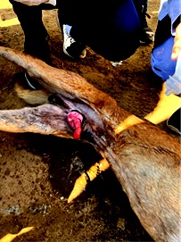
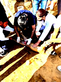
Fig: 1 Paraphimosis in dog Fig: 2 Application of topical lidocaine
History and Examination
A 3-year old male intact pariah dog was presented to the ambulatory unit of Apollo College of Veterinary Medicine, Jaipur, Rajasthan with the history of discomfort and a swollen extruded penis. Clinical examination revealed entrapment of the preputial skin behind the bulbus glandis with severe swelling of the bulbus glandis and penile tissue, and slight ulceration of the penile tissue with no evidence of ischemic necrosis of the penile tissue. Despite the edematous swelling at the base of penis, the general condition of dog was healthy and active. On the basis of clinical signs and physical examination, it was diagnosed as a case of paraphimosis.
Treatment and Management
Lidocaine gel 2% was applied topically over the penile tissue and the prepuce to reduce discomfort during manipulation for the reduction of the prepuce over the penis. The penis was gently cleansed to remove dirt with potassium permanganate (1:10000). The hyperosmolar paste of caster sugar (homemade granulated sugar) was applied on the swollen part of penis and prepuce to reduce the edema and swelling. The high solute concentration of caster sugar was used to osmotically draw out fluid from the edematous glans and foreskin prior to manual reduction (Fernández et al., 2001). Granulated sugar spread over the glans and foreskin for 30 min to 2 hours has been shown to reduce the swelling (Fernández et al., 2001). As the swelling subsided, an adequate lubricating agent, namely, paraffin wax was used for easy manipulation of the penis. The use of caster sugar (Bran) and paraffin combined with digital pressure facilitated the reduction and reposition of penis into the preputial sheath. After the reduction of the prepuce over the penis, a ring block using 2% lidocaine was injected subcutaneously in the preputial skin near the preputial orifice. The simple interrupted suture was placed ventrally in the preputial orifice using 2-0 silk suture to prevent reoccurrence. The dog was given cefotaxime @ 20 mg/kg and melonex @ 0.2 mg/kg intramuscularly for 3 days. The suture was removed after five days. Paraphimosis condition is more commonly seen in dog than cat and mostly in young dogs (Rochat, 2009). In this case, sexual hyperactivity associated with priapism could be the possible reason for paraphimosis as described earlier by Rao and Bharathi (2004) and Fossum (2007). However, several other factors including narrow preputial orifice, abnormal shortened penis, transmissible venereal tumour (TVT) and congenital causes may lead to paraphimosis (Fossum, 2007; Kumar, 2012). Recurrence of paraphimosis is common, especially when associated with sexual activity. To prevent recurrence, trimming of the preputial hair, careful inspection of the penis and prepuce after breeding, administration of progestogens and castration can be employed (Rochat, 2009). The combination of hyperosmolar solution with icepacks eased the return of exposed penis back into the preputial cavity (Elkins, 1984; Tiwari, 2004). The antibiotic (cefotaxime) administered in the present study was also effective as no secondary bacterial infection was noticed. After the treatment, the inflammatory swelling subsided and the animal recovered uneventfully with no recurrence up to three weeks of follow up.
REFERENCES
Boden, E. (2005). Black’s Veterinary Dictionary 21st edition Jaypee Brothers, Medical Publishers (P) Ltd. New Delhi. India. p. 512.
Davidson, A.P. (2020). Paraphimosis in Dogs and Cats. https://www.msdvetmanual.com
Elkins, A.D. (1984). Canine paraphimosis of unknown etiology. A case report. Veterinary Medicine. 79: 638-639.
Fossum, T.W. (2007). Surgery of the Reproductive and genital system. In: Small animal surgery, 3rd edition, Mosby Elsevier, pp: 768-770.
Kumar, A.; Sangwan, V.; Mahajan, S.K.; Singh, N.D.; Singh, K.; Anand, A. and Saini, N.S. (2012). Transmissible Venereal Tumor Induced Paraphimosis in Dogs. Journal of Advanced Veterinary Research, 2(1): 48-49.
Fernández, M.G.; Escandón, M.A.S.; Muntaner, L.P.; Pacios, J.C.L. (2001). Sugar: treatment of choice in irreducible paraphimosis. Actas Urol Esp.
Rochat, C.M., 2009. Paraphimosis and Priapism, Small Animal Critical Care Medicine, W.B. Saunders, pp. 615-618, https://doi.org/10.1016/B978-1-4160-2591-7.10141-9.
Rao, T.M and Bharathi, S. (2004). Paraphimosis associated with priapism in a dog. Blue Cross book. 22: 33-34.
Roberts, J.S. (1999). Infertility in male animals, pp. 648-649. In: Veterinary Obstetrics and Genital Diseases – 5th edition. CBS Publishers and Distributors, New Delhi.
Tiwari, S.K.; Sharda, R. and Dewangan, R. (2004). Successful Surgical Management of paraphimosis in a crossbred dog – A case report. Intas Polivet 5(2): 331-332.

